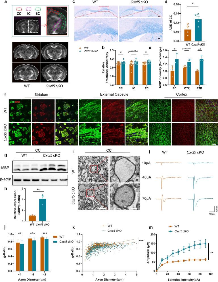
Fig. 3.
Astrocytic Cxcl5 deficiency alleviated BCAS-induced white matter damage. a, b Representative DTI axial views of FA maps (a) and quantification of FA values in the CC, IC and EC (b) acquired 2 months after BCAS. Two panels above show the areas (CC, IC and EC) of quantification. n = 12 per group. c, d Representative images of LFB staining (c) and quantification of AOD (d) in CC. Scale bar = 200 μm (top) and 50 μm (bottom). n = 4 per group. e, f Representative images for MBP (green) and NFH (red) double immunostaining (f) and quantification of the fluorescence intensity of MBP (e) in EC, CTX, STR. Scale bar = 50 μm. n = 3 per group. g, h Representative immunoblot bands of MBP (g) and quantification normalized to β-actin (h) in the CC. n = 3 per group. i–k TEM ultrastructural analyses of myelin integrity in the CC of WT and Cxcl5 cKO groups at 2 months after BCAS. i Representative TEM images in the CC. Scale bar = 5 μm (left) and 10 μm (right). j Comparison of the g-ratio of myelinated axons according to axon diameter between WT and Cxcl5 cKO mice. k Scatterplots of the myelin g-ratio as a function of the axon diameter. Axons were selected from 4 mice per group for measurement. n = 545 and n = 543 measured axons for each group. l, m Representative curves of CAPs of myelinated fibres (l) and quantification of the amplitude of evoked CAPs of myelinated fibres (m) in the CC. n = 3 mice and 10–12 recordings per group. All data were presented as the mean ± SEM. *p < 0.05, **p < 0.01, ***p < 0.001, ns means no significance. Student’s t-test for d, e, h. Mann–Whitney test for b, j, k. Two-way ANOVA with Bonferroni's post hoc test for m

