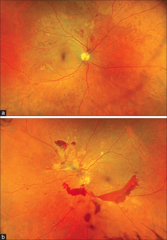
Figure 3.

Ultra-widefield fundus photograph (Optos California) of a patient with diabetic vitreous hemorrhage (Panel b). Fundus examination of the fellow eye shows proliferative diabetic retinopathy giving a clue toward etiology of vitreous hemorrhage (Panel a)
