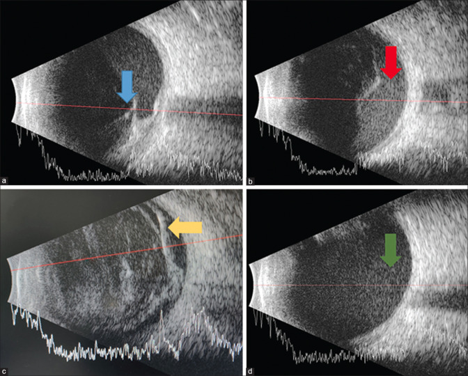
Figure 7.
Cross-sectional B-scan image with overlying A-scan of the eye showing tractional retinal detachment with associated sub-hyaloid hemorrhage (Panel a, Blue arrow), sub-hyaloid hemorrhage (Panel b, Red arrow), vitreous hemorrhage associated with retinal detachment seen as a double membrane (Panel c, Yellow arrow), and dispersed rebleed in the vitrectomized eye (Panel d, Green arrow)

