Figure 1:

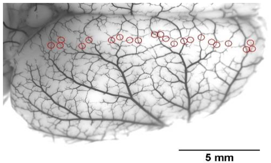
Photomicrograph of the dorsal surface of a rat brain perfused with India Ink to show leptomeningeal anastomoses (LMAs) or pial collaterals (red circles). Reproduced from Li Z et al., Am J Physiol 2018;315:H1703-H1712.

Photomicrograph of the dorsal surface of a rat brain perfused with India Ink to show leptomeningeal anastomoses (LMAs) or pial collaterals (red circles). Reproduced from Li Z et al., Am J Physiol 2018;315:H1703-H1712.