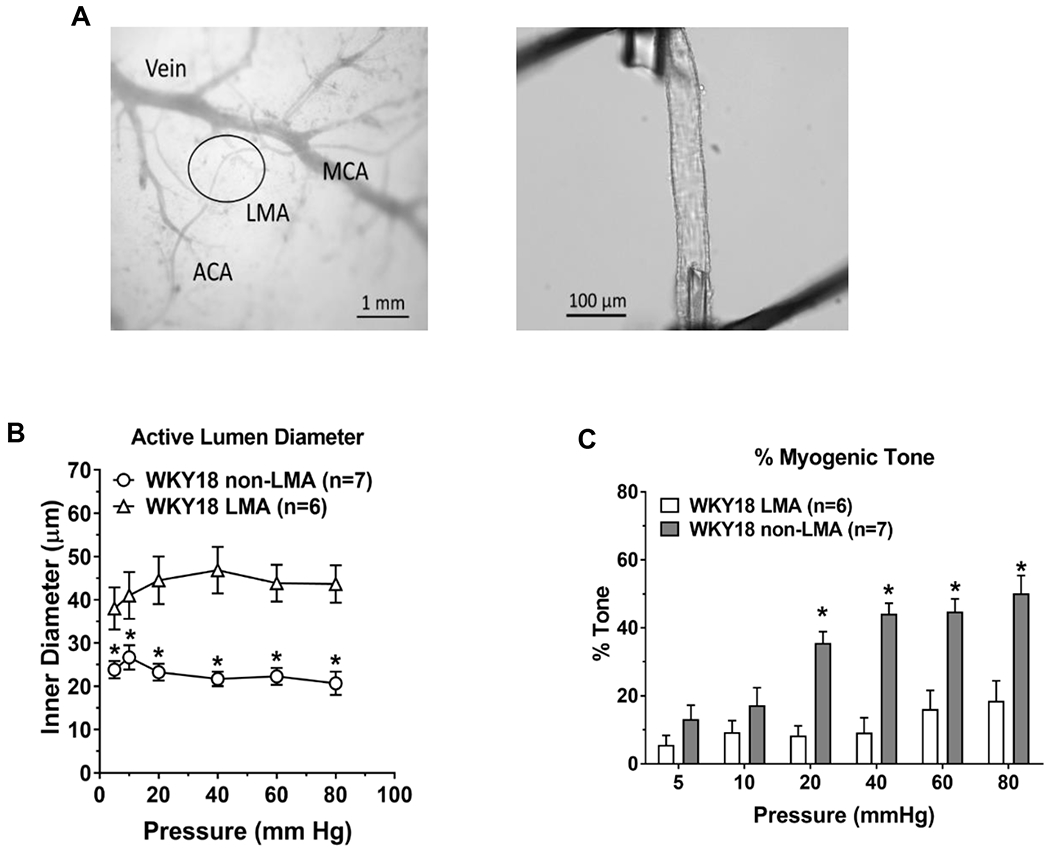
Figure 2:

The study of isolated and pressurized leptomeningeal anastomoses (LMAs) in an arteriograph system has allowed a greater understanding of the functional properties of these unique vessels. (A) Left, photomicrograph showing leptomeningeal arterioles (black circle) and how are identified as connecting distal branches of middle cerebral artery (MCA) and anterior cerebral artery (ACA) for study isolated and pressurized. Right, LMA isolated, mounted on glass cannulas within an arteriograph chamber and pressurized. Reproduced from Chan et al. Stroke 2016;47:1618-1625; Open access CC BY-NC-ND 4.0 B) Active myogenic vasoconstriction of LMAs and non-LMAs from 18 week old normotensive WKY rats. *P<0.05 vs. LMA. C) Myogenic tone of LMA and non-LMAs from 18 week old WKY rats. *P<0.05 vs. LMA. Reproduced from Cipolla MJ. Stroke 2021;52:2465-2477
