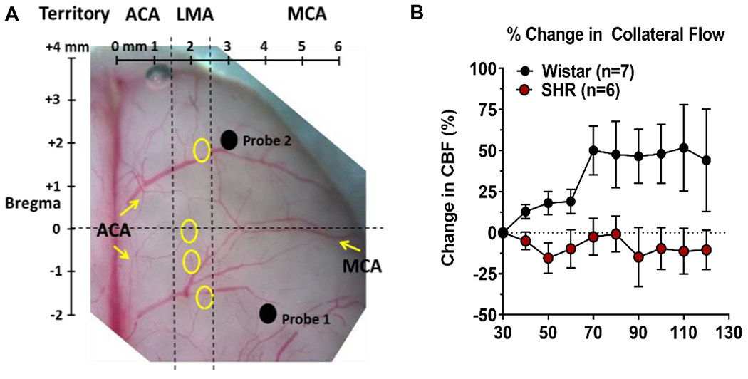
Figure 4:

Measurement of collateral flow using multi-site laser Doppler in rats and the effect of chronic hypertension. (A) Photomicrograph of the dorsal surface of a rat brain with overlay of skull coordinates. Probe 1 was placed over the core MCA territory and Probe 2 lateral to LMAs (yellow circles) to measure collateral flow. Reproduced from Cipolla et al., J Cerebr Blood Flow Metab. 2018;38:755-766. (B) Graph showing % change in collateral flow calculated from filament insertion in normotensive Wistar and hypertensive SHR rats. Collateral flow increased in response to MCAO in Wistar but not SHR. Data will be made available upon reasonable request.
