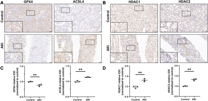
FIGURE 1.
The expression of GPX4, ACSL4, HDAC1 and HDAC2 in acute kidney injury from human biopsies. Representative images of immunohistochemistry of GPX4 and ACSL4 (A), HDAC1 and HDAC2 (B) in the healthy control and AKI human kidney biopsy specimens. Scale bar = 50 μm. Quantification of examining positive staining area of GPX4 and ACSL4 (C), HDAC1 and HDAC2 (D) in tubulointerstitium normalized to healthy control (Non-tumor kidney tissue from the patients who had renal cell carcinoma and underwent nephrectomy was used as control). n = 3. ✱✱ value p < 0.01. Statistically analyzed via a one-way ANOVA with Dunnett’s correction.

