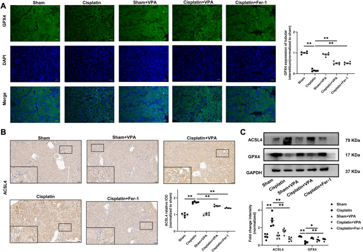
FIGURE 3.
VPA treatment attenuates expression of ferroptosis-related proteins in cisplatin-induced AKI mice. Representative images of immunofluorescence of GPX4 in the tubulointerstitium of sham, cisplatin and VPA or Fer-1 treatment groups. Quantification of examining positive staining area of GPX4 in the tubulointerstitium normalized to sham group (A). Immunohistochemical staining and semi-quantitative analysis for expression of ACSL4 (B) in different groups as indicated. Representative images of Western blot from different groups as indicated, including ACSL4 and GPX4, respectively (C). Scale bar = 50 μm. n = 5. ✱✱ value p < 0.01, ✱ value p < 0.05. Statistically analyzed via a one-way ANOVA.

