Figure 3.
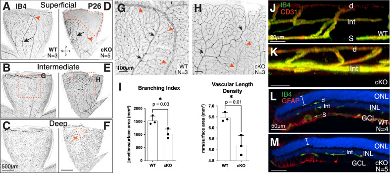
Tbx3 cKO retina failed to form superficial plexus in the adult dorsal retina. (A–F) Confocal flatmount images of P26 dorsal retinas from WT (A–C) and cKO (D–F) mice stained with isolectin GS-B4 (IB4). Confocal images are inverted. The superficial (A, D), intermediate (B, E), and deep (C, F) plexus from the same retinal flatmounts are shown. (G, H) Maximal projection images from a stereomicroscope simultaneously showing all three plexi from the region indicated in B and E. Arteries (black arrows) and veins (red arrowheads) in each panel are marked. (I) Branching index (branch points/retinal surface area) and vascular length density (vessel length/retinal surface area) from maximum projections images (see Supplementary Fig. S2 for quantitation). Statistical significance was determined using an unpaired, two-tailed t-test; P values are shown on the graph. (J, K) Still, three-dimensional, rendered confocal images of retinal plexus in dorsal retina of WT (J) and cKO (K) mice stained with IB4 (green) and anti-CD31 antibody (red). (L, M) Transverse cryostat sections of P26 retina stained with IB4 (green) and anti-GFAP antibody (red) to mark endothelial cells and astrocytes, respectively. The superficial vascular plexus (S), intermediate vascular plexus (Int), and deep vascular plexus (d) are marked; the outer nuclear layer (ONL), inner nuclear layer (INL), and GCL can be seen with the nuclei marker DAPI (blue). Note that the anti-GFAP (raised in mouse) antibody lightly stained the vascular cells, as these mice were not perfused.

