Figure 6.
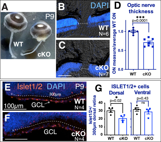
Dorsal-specific loss of RGCs with loss of Tbx3. (A) Snapshot of whole eyes with optic nerves from P9 WT and cKO siblings. (B, C) Sections of WT and cKO eyes stained with nuclei marker (DAPI) to visualize neural retina. The dashed line indicates optic nerve thickness in each sample. (D) Graph quantitating the optic nerve thickness in WT and cKO eyes. (E, F) Transverse cryostat sections of P9 WT and cKO dorsal tip immunostained with anti-Islet1/2 antibody. The numbers of Islet1/2-positive cells in the first 300 µm of the most dorsal and ventral retina were counted (dashed line). (G) Graphs of the Islet1/2-positive cells in the dorsal and ventral regions. Statistical significance was measured using an unpaired, two-tailed t-test for the graphs in D and G.

