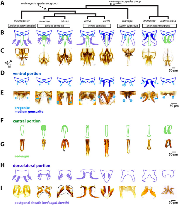
Figure 2: The rapidly evolving phallus is composed of three main components.
A) Phylogeny for eight species of the melanogaster species group based on Obbard et al., 2012 with nodes that contain the melanogaster species subgroup and melanogaster species group indicated by arrows. B) A schematic breakdown of the adult phalluses of each species. C) Light microscopy images of the whole adult phallus for each species. Image stacks that show the relative position of each part can be found in Supplementary videos. D) Schematic representation of the ventral portion of the phallus (dark blue) which contains the pregonites (light blue) and processes (filled in dark blue) in D. erecta and D. ananassae. Light blue asterisks designate the position of the pregonites. E, G, I: Light microscopy images of microdissections of the adult phallus (here in lateral view, distal end pointing downward) separated in the ventral, central, and dorsolateral portions. E) Microdissections of the ventral portion processes shows the processes of D. erecta and D. ananassae are connected to the pregonites. Light blue asterisks designate the position of the pregonites. F) Schematic representation of the central portion of the phallus (light green), which contains processes (filled in light green) in D. santomea, D. teissieri, D. orena, and D. malerkotliana. G) Microdissections of the aedeagus confirm that the processes are physically attached to aedeagus. The aedeagus of the D. malerkotliana is translucent (dashed line) with only the process sclerotized. H) Schematic representation of the dorsolateral portion of the phallus (light purple), which contains two pairs of processes (filled in light purple) in D. melanogaster, and one pair in D. santomea, D. teissieri, D. erecta, and D. orena. I) Microdissection confirms that the processes are physically attached to postgonal sheath. In D. santomea, D. teissieri, D. erecta, and D. malerkotliana portions of the anterior postgonal sheath are translucent (outlined with dashed lines). The image of the D. orena dorsolateral portion was created by copying and mirroring one side of the structure, as it was difficult to flatten intact for imaging. Aedeagal sheath is an alternative term for postgonal sheath.

