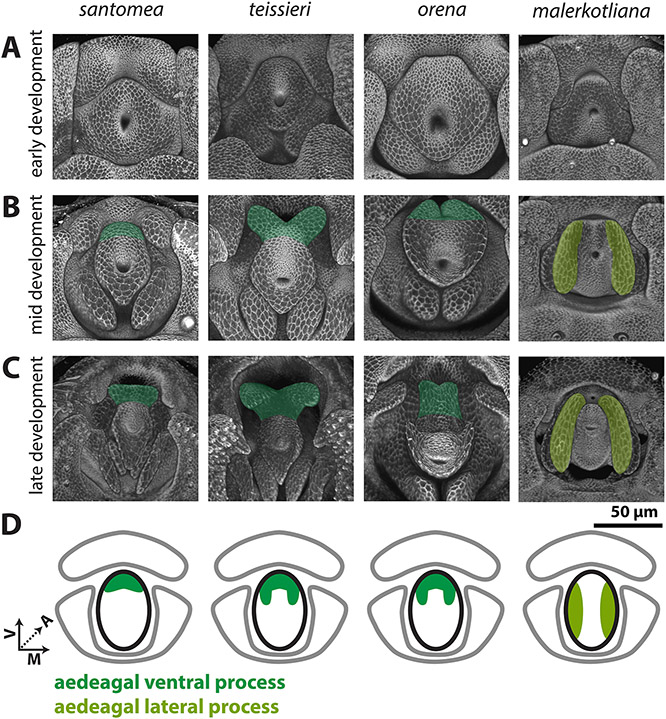
Figure 5: Processes developing from the central primordium are found in the yakuba/erecta and bipectinata complexes.
A-C) Signal from the apical cellular junctions (ECAD) was used to highlight the overall morphology of developing pupal genitalia. A) Early in development, the central primordium forms a flat donut-shaped structure in all species shown. B) By mid-development, the ventral portion of the aedeagus is extended in D. santomea, D. teissieri, and D. orena in what will form the aedeagal ventral process (dark green). In D. malerkotliana the lateral edges of the central primordium extend anteriorly in what will form the aedeagal lateral process (yellow-green). C) By late development, the aedeagal ventral process and the aedeagal lateral process further extend from the aedeagus. D) Schematic representations of the aedeagal ventral process (dark green) and aedeagal lateral process (yellow-green) showing where they connect to the aedeagus (outlined in black). All images have the same axes, V (Ventral), A (Anterior), M (Medial) and are the same scale.

