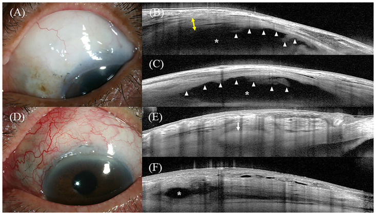
Fig 5. Representative images of AS-OCT.
Fig A-C represent a successful case and Fig D-F represent a failed case. (A) Slit-lamp photo of the bleb at 19 months after trabeculectomy with AMT. The IOP preoperatively was 21 mmHg and at the time of the AS-OCT examination was 7 mmHg. (B) Vertical AS-OCT scan of the bleb. The AS-OCT showed a tall bleb with a hyporeflective striping layer (yellow two-way arrows). Fluid-filled space (asterisk) extended posteriorly beyond the view of the image field. Amniotic membrane was noted even >19 months after surgery (white arrow head). (C) Horizontal AS-OCT scan of the bleb. The AS-OCT showed a diffuse fluid-filled space with the amniotic membrane (white arrow head). (D) Slit-lamp photo of the bleb at 6 months after trabeculectomy with AMT. The IOP preoperatively was 23 mmHg and at the time of the AS-OCT examination was 19 mmHg. (E) Vertical and (F) Horizontal AS-OCT scans of the bleb. The white arrow indicates the scleral flap. The bleb height was low, with limited fluid-filled space (asterisk) and no striping layer. AS-OCT, anterior segment optical coherence tomography; AMT, amniotic membrane transplantation; IOP, intraocular pressure.

