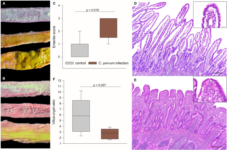
Figure 2.
Macroscopic and histopathologic findings. Macroscopic differences in the consistency of the ingesta in cranial (top) and caudal small intestine (middle) and colon (bottom) of A control calves and B C. parvum-infected calves are most obvious in the colon. C Macroscopic evaluation revealed a severe, acute to subacute, diffuse, catarrhal enteritis in the C. parvum-infected calves (red boxes), whereas all but one calves of the control group (gray boxes) displayed no enteritis; N = 5, Mann-Whitney Rank Sum Test, p < 0.05. The main histopathological finding is a marked reduction in the small intestinal villus length in the C. parvum-infected (D) as compared to control calves (E); scale bar = 200 μm, hematoxylin-eosin staining. Higher magnification (inserts) displays multiple apicomplexan cysts characteristic of C. parvum; scale bar = 20 μm. F Marked villus atrophy as the histopathological correlate of the macroscopic enteritis is demonstrated by a decreased villus-crypt-ratio in the jejunum of infected calves (red boxes) compared to control calves (gray boxes). N = 5, Student’s t-test. Boxes show median and percentiles plus error bars.

