
Figure 1. Placental anatomy and parenchymal lesions:
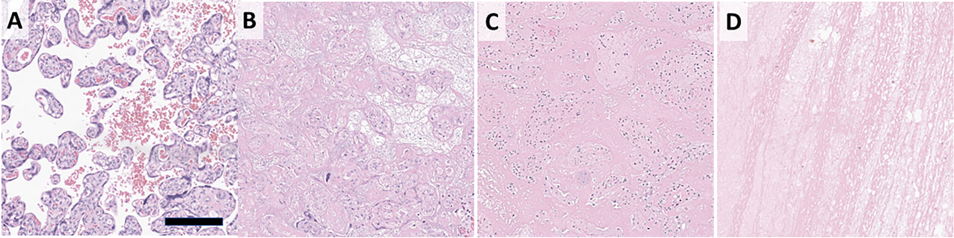
Normal placental histology showing villi are bathed in maternal blood (A). Infarctions are due to loss of maternal blood flow and characterized by indistinct villi that are either back-to-back or with loose material between (B). PVFD shows loss of syncytiotrophoblast layer from the villous surface with replacement of the intervillous space by dense pink fibrin (C). IVT displace villi and show characteristic lines of Zahn (D). Scale bar 200 μm.