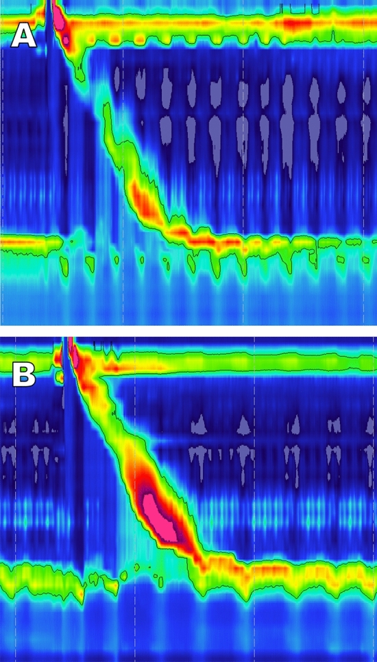
Fig. 2.

HRM topographic plot before (A) and after MSA implantation (B), but before removal in a patient who subsequently underwent device removal for dysphagia. At baseline (A) the patient has adequate distal contractile integral (DCI) with complete bolus clearance (not shown). There is complete LES relaxation and evidence of some degree of crural-LES separation, indicating a hiatal hernia. Following device implantation (B) the topographic plot shows an elevated intrabolus pressure (iBP) and integrated relaxation pressure (IRP) and a compensatory increase in esophageal body contractility
