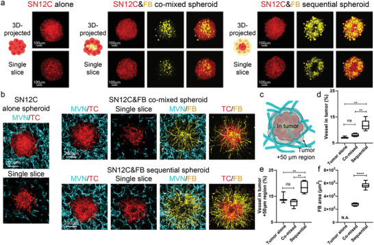
Figure 2.

Sequential SN12C tumor spheroid shows the highest vascularization level. a) Schematic, 3D‐projected image, and single slice image of SN12C tumor spheroids formed by tumor alone, co‐mixed, or sequential methods. b) 3D‐projected images of vascularized tumor spheroid formed by tumor alone, co‐mixed, or sequential methods. TC is short for tumor cells. c) Schematic showing the tumor region and nearby 50 µm distance region for vascularization analysis. d) Statistical analysis of vessel percentage in the tumor region or e) tumor +50 µm region. f) Area analysis of FBs from tumor spheroid. Bars represent mean ± S.D. One‐way ANOVA was performed for the statistical comparison in (d) (p < 0.001) and (e) (p < 0.001). Significance was determined using Tukey's multiple comparisons test of mean values between each group. Two‐tailed t tests were performed for the statistical comparisons in figure (f). Data were collected from at least 6 tumor spheroids for each group. *p < 0.05, **p < 0.01, ***p < 0.001, ****p < 0.0001.
