Fig. 1.
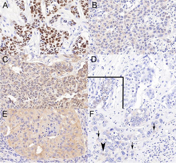
Examples of TRPS1 positive (ABC) and negative (DEF) immunostainings. A Obvious positive staining with strong nuclear labelling and no cytoplasmic background. B, C Weak nuclear staining in > 10% of the cells interpreted as positive or negative by 50% of the observers; note the relatively strong background cytoplasmic staining. D Completely negative reaction, inset showing positive staining in a normal duct of the same TMA core. E A case with strong cytoplasmic background staining ignored due to the lack of nuclear staining. F The case with about 5% of the nuclei staining weakly (bold arrowhead) or in a barely visible fashion (arrows). All ×40, TRPS1

