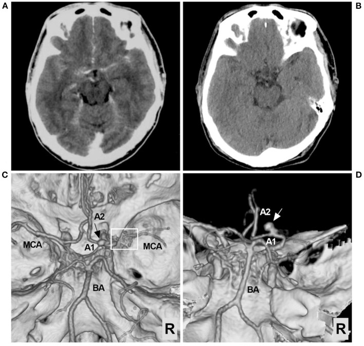
Figure 1.
Preoperative CT and CTA images. (A) CT showing the extensive SAH, focusing on the left suprasellar cistern and the base of the Sylvian fissure. (B) Six days later, repeat CT showing absorption of the SAH. (C) CTA of the superior-inferior view showing the arterial network of the right proximal MCA (frame) and an aneurysm (arrow). (D) CTA of the posterior-anterior view showing the aneurysm in detail; the aneurysm (arrow) was located in the A1 segment of the ACA. ACA, anterior cerebral artery; A1, first segment of the ACA; A2, second segment of the ACA; BA, basilar artery; CT, computed tomography; CTA, CT angiography; MCA, middle cerebral artery; R, right; SAH, subarachnoid hemorrhage.

