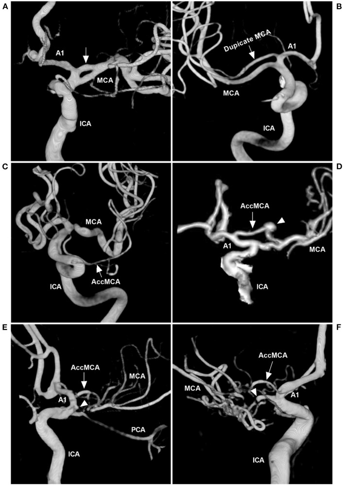
Figure 4.
MCA anomalies on angiography. (A) Three-dimensional DSA of the ICA showing a fenestration (arrow) at the beginning of the MCA. (B) Three-dimensional DSA showing a duplicate MCA (arrow). (C) Three-dimensional DSA showing a type 1 AccMCA (arrow) from the ICA trunk. (D) Three-dimensional DSA showing a type 2 AccMCA (arrow) with complete anastomosis between the A1 middle segment and MCA forming a large fenestration; the arrowhead indicates an aneurysm. (E) Three-dimensional DSA showing a twig-like MCA supplied by the AccMCA (arrow) from the A1 end; the MCA did not supply the twig-like MCA, and the MCA origin presented with a protrusion (arrowhead). (F) Three-dimensional DSA showing that the twig-like MCA was supplied by the AccMCA from the A1 origin (arrow); the MCA supplied the twig-like MCA (arrowhead). A1, first segment; AccMCA, accessory middle cerebral artery; CT, computed tomography; DSA, digital subtraction angiography; ICA, internal carotid artery; MCA, middle cerebral artery; PCA, posterior cerebral artery.

