. 2023 May 2;11:1115. Originally published 2022 Sep 28. [Version 3] doi: 10.12688/f1000research.125645.3

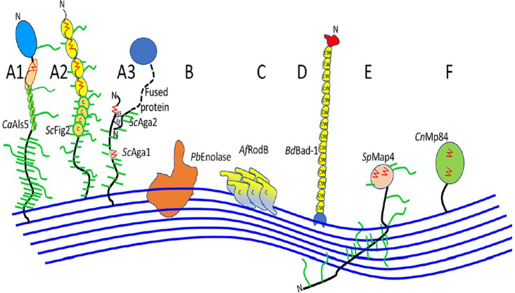
Figure 3. An illustration of cell wall association in fungal adhesins (the blue line indicates the cell wall which is composed of glucan polymers; Ca, C. albicans; Sc, S. cerevisiae; Pb, P. braziliensis; Af, A. fumigatus; Bd, B. dermatitidis; Sp, S. pombe; Cn, C. neoformans; A, adhesins; B-E, modes of cell wall attachment; A1 and A3, have discrete ligand binding domains; A2 doesn’t have discrete ligand binding domains, Cys-rich sequences in ScFig2; C, Cys-rich sequences in AfRodB; D, Cys/Trp rich domains in Bd/Bad-1; F, attached by modified GPI anchors to the cell wall) ( Lipke 2018).

Figure 3.