. 2023 Apr 25;24(5):406–423. doi: 10.3348/kjr.2022.0998

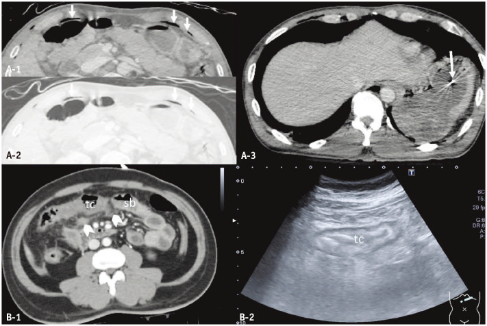
Fig. 10. False positives of surgical bowel injuries. A: Pseudopneumoperitoneum (arrows) and bullet fragment (long arrow) in the gastric lumen of a 44-year-old man who sustained multiple gunshot wounds to the face, neck, and shoulder. B: Blunt non-surgical colonic and mesenteric injuries in a 51-year-old man. Axial computed tomography (CT) image (A-1) demonstrates curvilinear streaks of air (arrows) in the anterior aspect of the abdomen that closely follow the inner aspect of costal cartilages. They demonstrate internal reticular strands in the lung window setting (A-2), and are continuous with chest wall emphysema (not shown). Axial CT image (A-3) reveals a bullet fragment (long arrow) in the gastric lumen, which was presumably retained in the mouth then ingested as there is no entry or exit wounds in the torso. Axial CT (B-1) and ultrasound (B-2) images demonstrate thickening and decreased enhancement of the transverse colon (tc), and fat stranding with small hematoma (arrowheads) of the transverse mesocolon. However, the diagnostic laparoscopy does not demonstrate full-thickness injury or bowel ischemia. sb = small bowel.

Fig. 10