. 2023 Apr 25;24(5):406–423. doi: 10.3348/kjr.2022.0998

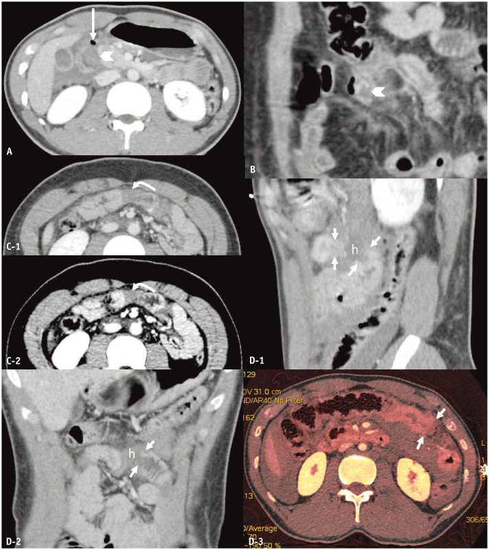
Fig. 2. Bowel wall defect in full-thickness bowel tears. A: Blunt duodenal trauma in a 20-year-old man. B–D: Blunt jejunal trauma in a 44-year-old man (B), a 22-year-old man (C), and a 27-year-old man (D). Axial (A) and coronal-reformatted (B) computed tomography (CT) images indicate incomplete rings of enhancement (arrowheads pointing at the defect) of the D2 segment of duodenum and jejunum, respectively, with wall thickening. A long arrow points at an extraluminal air bubble. Axial CT images in standard (C-1) and narrow (C-2) window width settings reveal a Janus sign (curved arrows) of the jejunum in the mid-abdomen. Sagittal-reformatted (D-1), coronal-reformatted CT images (D-2), and axial iodine map (D-3) demonstrate a cutoff or discontinuation (short arrows) of the jejunal loop in the left upper quadrant separated by a hematoma (h). Long arrow = pneumoperitoneum.

Fig. 2