. 2023 Apr 25;24(5):406–423. doi: 10.3348/kjr.2022.0998

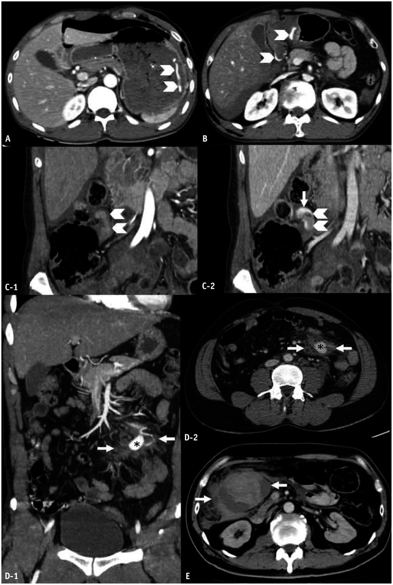
Fig. 3. Active vascular contrast extravasation (AVCE), pseudoaneurysm, and hematoma of the mesentery. A: Stab gastric trauma in a 30-year-old man. B–E: Blunt mesenteric trauma in a 44-year-old man (B), a 45-year-old man (C), a 36-year-old woman, (D) and a 49-year-old man (E). Axial computed tomography (CT) images (A, B) reveal AVCE as irregular linear contrast jets (arrowheads) within the gastric lumen (A) and within a hematoma of the gastroduodenohepatic ligament (B). Coronal-reformatted CT images in arterial (C-1) and portovenous (C-2) phases indicate an AVCE (arrow) as an enhancing hematoma (arrowheads). Coronal-reformatted, arterial-phase (D-1), and axial portovenous-phase (D-2) CT images demonstrate an oval pseudoaneurysm (asterisks) arising from the jejunal branch of the superior mesenteric artery, which is surrounded by a hematoma (arrows). The axial CT image (E) depicts a large mixed-attenuation hematoma (arrows) of the ascending colonic mesentery without AVCE.

Fig. 3