. 2023 Apr 25;24(5):406–423. doi: 10.3348/kjr.2022.0998

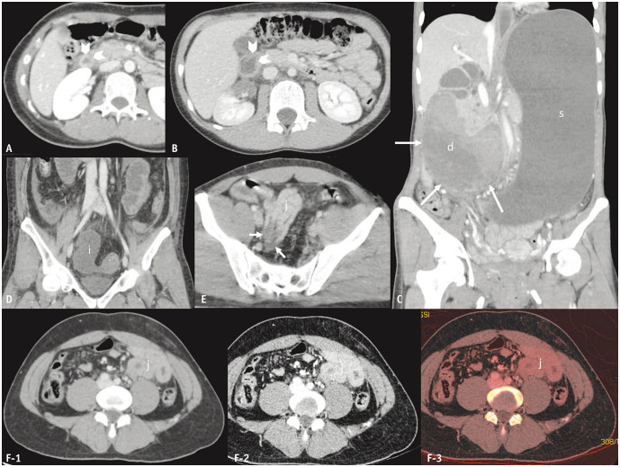
Fig. 4. Thickening, hematoma, and abnormal enhancement of the bowel walls. A–C: Blunt non-perforated duodenal trauma in three female patients aged 20, 10, and 36 years, respectively. D: Blunt ileal trauma in a 45-year-old man with a bucket-handle mesenteric injury and ileal ischemia. E, F: Blunt jejunal trauma (full-thickness tear) in a 29-year-old (E) and a 43-year-old man (F). Axial (A, B) computed tomography (CT) images indicate wall thickening (arrowheads) of the duodenum that is circumferential and uniform (A) and non-uniform (B) surrounded by periduodenal fluid. Coronal-reformatted CT image (C) demonstrating a large, obstructive, heterogeneous hematoma (long arrows) in segments D2 and D3 of the duodenum (d). The coronal-reformatted CT image of another patient (D) depicts absent wall enhancement of the fluid-filled dilated ileal loop (i). A segmental decrease in enhancement (short arrows) of the collapsed jejunum (j) was observed in another patient (E). Axial CT images with standard (F-1) dual-energy setting reveal a segment of increased jejunal wall enhancement (j), although the findings are more readily visible on the low-keV setting (F-2) and iodine map (F-3). d = duodenum, i = ileum, j = jejunum, s = stomach.

Fig. 4