. 2023 Apr 25;24(5):406–423. doi: 10.3348/kjr.2022.0998

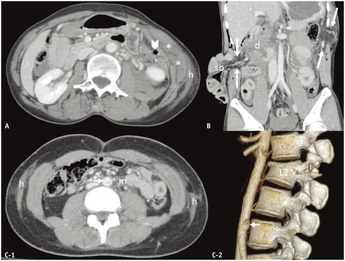
Fig. 7. Abdominal wall injuries and seat belt syndrome. A: Abdominal wall hemorrhage in a 58-year-old woman with a full-thickness colonic tear. B: Abdominal wall muscular tears and bowel evisceration in a 28-year-old man with multiple sites of full-thickness bowel tears. C: Seat belt syndrome in a 25-year-old woman with mesenteric hemorrhage (m), infrarenal abdominal aortic pseudoaneurysm (arrow), and flexion–distraction injury of the L2 vertebra. Axial computed tomography (CT) images (A, C-1) demonstrate subcutaneous hemorrhages (h) as well as enlargement and heterogeneous attenuation of the obliques and transversus abdominis muscles with fascial hemorrhage (asterisks). Coronal-reformatted CT image (B) depicts two defects of the abdominal wall (between long arrows): one at the right lateral abdominal wall muscles through the muscles, subcutaneous fat, and skin resulting in evisceration (sb), and another through the left intercostal muscle, resulting in herniation of omental fat. Volume-rendered 3D CT image (C-2) displays an infrarenal aortic pseudoaneurysm (arrow) and an L2 flexion–distraction injury. Arrowhead = pneumoperitoneum, d = cutoff of duodenum, L2 = second lumbar vertebra, m = mesenteric hematoma, sb = small bowel.

Fig. 7