. 2023 Apr 25;24(5):406–423. doi: 10.3348/kjr.2022.0998

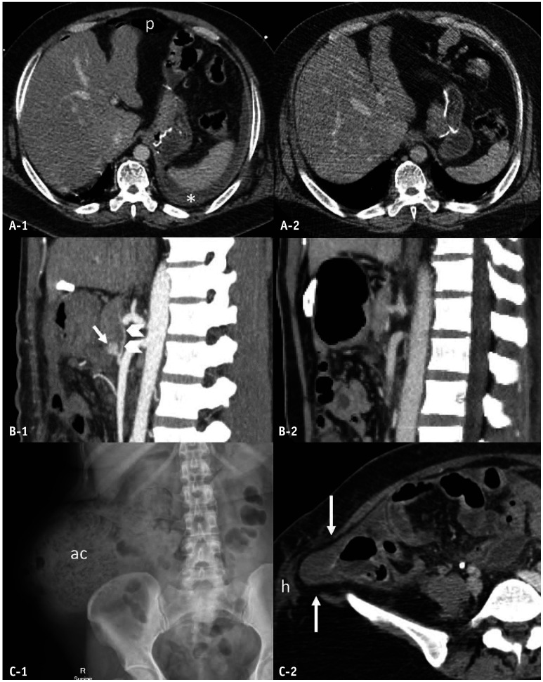
Fig. 9. Delayed presentation of surgical bowel and mesenteric injuries. A: Blunt gastric trauma (full-thickness tear) in a 43-year-old man with a history of gastric bypass surgery. B: Blunt traumatic pseudoaneurysm (arrow) of the superior pancreaticoduodenal artery (SPDA; arrowheads) in a 36-year-old woman with delayed onset of lower gastrointestinal hemorrhage following repair of traumatic duodenal perforation. C: Traumatic lumbar hernia in a 44-year-old woman. Axial computed tomography (CT) image obtained 26 h after trauma (A-1) indicates free fluid (asterisk) and pneumoperitoneum (p) secondary to full-thickness gastric tear, not observed in the initial CT (A-2). Sagittal-reformatted maximal intensity projection CT (B-1) performed 16 days after trauma demonstrates an eccentric outpouching pseudoaneurysm (arrow) of the SPDA (arrowheads), not visualized on the initial post-laparotomy CT (B-2). Conventional radiography of the abdomen performed 9 years after trauma (C-1) reveals a large, ascending-colon-containing lumbar hernia (ac). The initial CT at the time of trauma (C-2) reveals a defect of the right posterolateral abdominal wall muscle (long arrows) with subcutaneous hemorrhage (h). ac = ascending colon.

Fig. 9