-
Ai
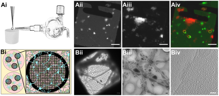
Glass atomizer used to disperse depolarizing solution on the EM grid milliseconds before the grid is vitrified.
-
Aii
Spray droplets imaged with the GFP filter set. Scale bars, 20 μm.
-
Aiii
Synaptosomes imaged with the DAPI filter set. Scale bar, 20 μm.
-
Aiv
Overlay of spray droplets (green) and synaptosomes (red). Yellow circles show contact between droplets and synaptosomes in thin area, which is suitable for cryo‐ET. Scale bar, 20 μm.
-
Bi
Schematic drawing of a six‐well Petri dish depicting astrocytes (pink) growing at the bottom of the Petri dish below EM grids (black, round grid overlaying the astrocytes), with neurons (blue) growing on top of the grids.
-
Bii
Grid square overview with neurons growing over it. Scale bar, 5 μm.
-
Biii
Medium‐magnification overview of neurons growing over R2/1 holes. Scale bar, 500 nm.
-
Biv
One slice of a tomogram depicting a synapse. Scale bar, 100 nm.

