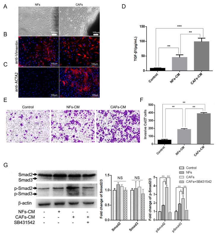
Figure 1 .
CAFs promote OSCC invasion through the TGF-β1/Smad2/3 pathway
(A) NFs were derived from normal tongue tissue. CAFs were derived from tongue carcinoma. (B) NFs and CAFs were both positive for vimentin (red). (C) CAFs were positive for ACTA2 (red), whereas NFs were not. (D) The amount of secreted TGF-β1 from control cells, NFs and CAFs after 24 h was detected. (E) Crystal violet staining of invaded Cal27 cells in control, NF-CM and CAF-CM. (F) Histogram showing that CAFs prominently promoted Cal27 cell invasion. (G) Western blot analysis of Smad2/3 and p-Smad2/3 proteins in Cal27 cells with different stimulations by NF-CM, CAF-CM or CAF-CM with SB431542. * P<0.01, ** P<0.001 and *** P<0.0001. Scale bar: 100 μm. Data are presented the mean±standard deviation.

