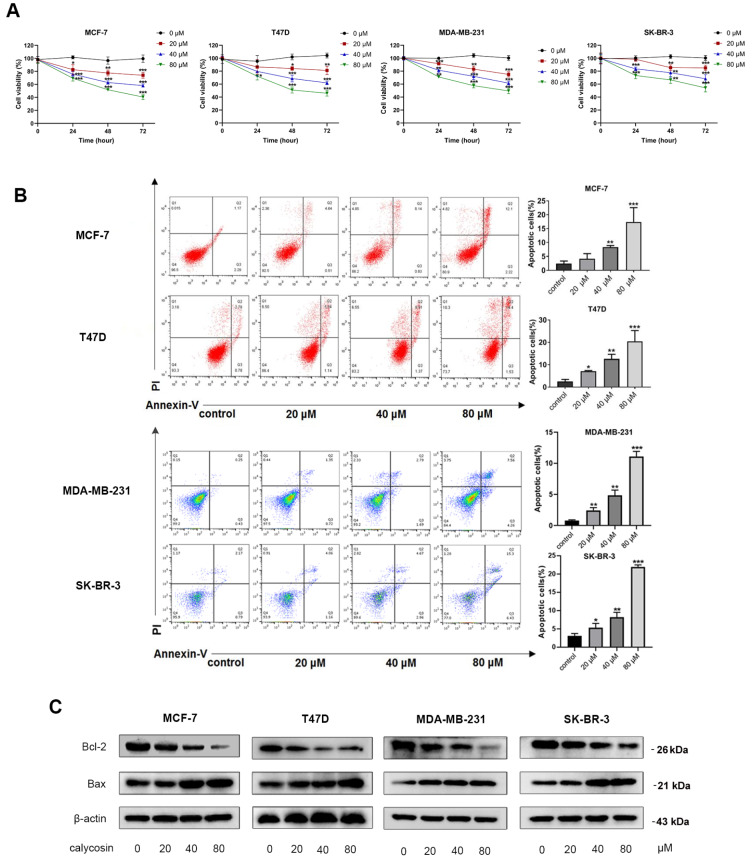
Figure 1 .
Inhibition of calycosin in breast cancer cell progression in both ER-positive and ER-negative breast cancer cell lines
Different concentrations of calycosin (0, 20, 40 and 80 μM) were used to treat ER-positive breast cancer cells (MCF-7 and T47D) and ER-negative breast cancer cells (MDA-MB-231 and SK-BR-3). (A) The viability of cells was detected by CCK-8 assays. (B) Flow cytometry was performed to detect the apoptotic rate after 48 h of calycosin treatment. (C) Western blot analysis was applied to detect the protein expression levels of Bcl-2 and Bax after 48 h of calycosin treatment. β-Actin was used as an internal control. Data are presented as the mean±SD ( n=3). * P<0.05, ** P<0.01 and *** P<0.001 compared with the control group.

