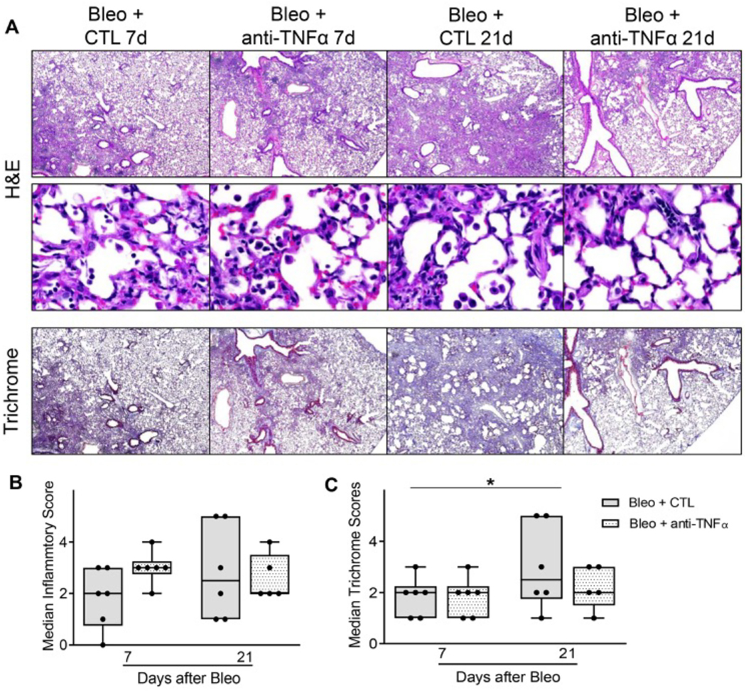
Fig. 2.
Effects of anti-TNFα antibody on bleomycin-induced lung histolopathology and cell counts. Panel A: Hematoxylin and Eosin and Masson’s Trichrome stain of tissue sections were prepared 7 d, and 21 d after exposure of mice to bleomycin (Bleo + CTL) or Bleo + anti-TNFα antibody. Original magnification: 40× (top and bottom panels) and 400× (middle panels). Representative images from at least 3 mice/treatment group are shown. Panels B and C: Histopathological scoring of sections was based on qualitative score of perivascular and peribronchial infiltrate, edema, number and extent of injury foci, alveolar thickening. Data are represented as median scores. *Significantly different (p ≤ 0.05) from Bleo + CTL group.

