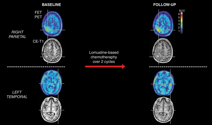
Figure 6.
Patient with a right parietal glioblastoma with an unfavorable survival (patient #22). After two cycles of lomustine chemotherapy, the contrast-enhancing lesion on MRI appeared unchanged (Stable Disease according to RANO criteria) compared to the baseline MRI scan. In contrast, the corresponding FET PET at follow-up showed, relative to the baseline scan, an increased maximum tumor-to-brain ratio (TBRmax) and metabolic tumor volume (MTV) (relative increase, 21% and 14%, respectively). Additionally, a new distant hotspot lesion at follow-up was observed in the left temporal lobe on FET PET but not MRI. The patient had an unfavorable outcome with a PFS of 1.7 months and an OS of 9.1 months.

