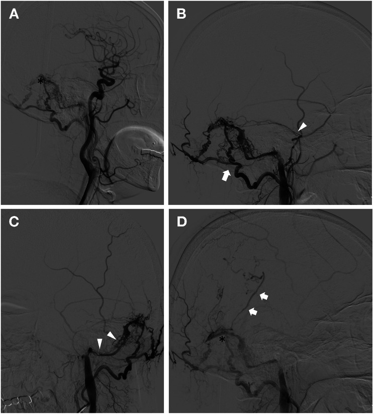
Fig. 2.
Cerebral digital subtraction angiography of the patients. (A) The Cognard type IIb dural arteriovenous fistula in the transverse sinus (black asterisk) is observed on digital subtraction images of a cerebral angiogram after left common carotid artery contrast injection. (B) The fistula was filled via shunts fed by the left occipital artery (white arrow) and branches of the middle meningeal artery (white arrowhead). (C) The downstream left sigmoid sinus (white arrowhead) appeared severe stenosis. (D) Retrograde venous drainage from the left transverse sinus (black asterisk) back into the vein of Labbe (white arrow) was observed during the late phase of cerebral angiogram

