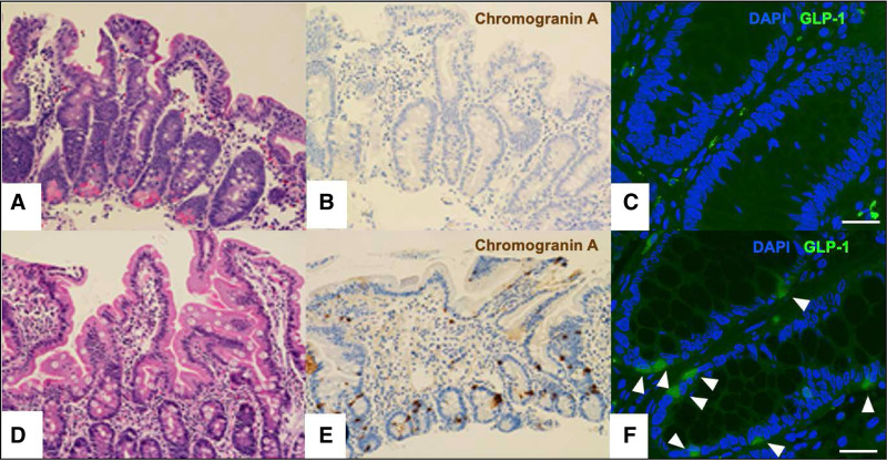
FIGURE 1.
Intestinal biopsies demonstrate a paucity of enteroendocrine cells as stained using Chromogranin-A and GLP-1. Patient (A) duodenum with H&E stain (200×), (B) duodenum with Chromogranin A stain (200×), and (C) colon with GLP-1 immunofluorescent stain (scale bar 25 μm). Age-matched control (D) duodenum with H&E stain (200×), (E) duodenum with Chromogranin A stain (200×), and (F) colon with GLP-1 immunofluorescent stain highlighting GLP-1 expressing enteroendocrine cells (arrowheads, scale bar 25 μm).

