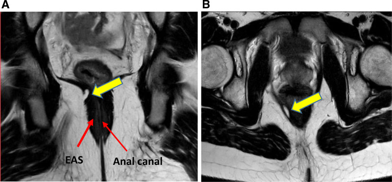
FIGURE 1.
Pelvic MRI showing: (A) T2-coronal oblique and (B) T2-axial views. MRI = magnetic resonance imaging. The yellow arrows show the fat protrusion through the external anal sphincter (EAS) defect reported only at 8 o’clock (lithotomy position). The anal canal in collapsed in the midline (figure 1A) with no abnormality identified in the internal anal sphincter (IAS), (thin wall lining the anal canal).

