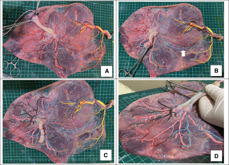
Figure 2.
Placental perfusion staining. (A) In the 1st step, the umbilical veins of both fetuses were perfused with yellow dye and pink dye, respectively. (B) In the 2nd step, 1 umbilical artery of the smaller fetus was perfused with blue dye, and blue staining of the branch vessels of the 2 umbilical arteries of the smaller fetus and 1 umbilical artery of the larger fetus through the arterial-arterial anastomosis (white arrow) was observed. (C) In the 3rd step, the other umbilical artery of the larger fetus was perfused with green dye. (D) No color mixing was observed between the 2 umbilical arteries of the larger fetus and their branch vessels).

