
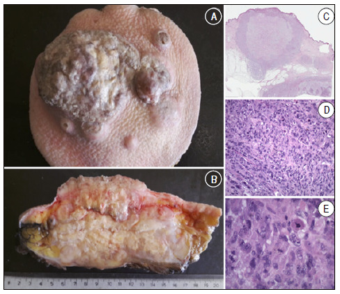
Figure 4. A and B: Macroscopy; soft tissue tumor in the hip, surface with ulcerated center. Affects a deep margin; C, D and E: Microscopy (H&E) revealing cytological and nuclear pleomorphism. The tumors often contain giant cells, spindle cells, and histiocyte-like round cells in varying proportions. Storiform cell patterns and chronic inflammatory stroma are also common. The spindle component resembles fibroblasts, myofibroblasts, or smooth muscle patterns. ( 4 .
Source: A cortesy from pathology IOT HC FMUSP/CEDAP Joinville.