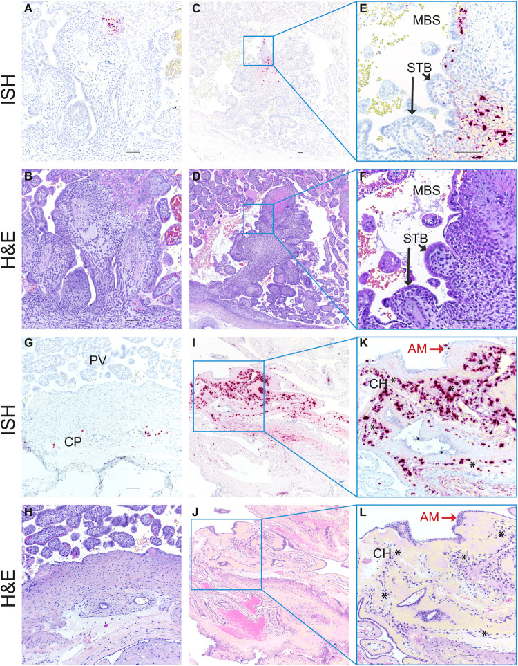
Fig 6. ZIKV RNA detected in MFI by in situ hybridization (ISH).
Photomicrographs of paraffin embedded sections of the placenta and fetal membranes from 45/14-4. Panels A, C, E, G I, and K Positive ISH indicated by the red/pink chromogenic stain. Panels B, D, F, H, J, and L are corresponding H&E-stained sections to provide histological detail. Panels E, F K and J are higher magnification images of the corresponding regions in the adjacent photomicrographs. Scale bar represents 100 μm. Abbreviations: MBS maternal blood space, STB syncytiotrophoblasts, CP chorionic plate, PV placental villi, AM amniotic membrane, CH chorionic membrane. Black arrows indicate STB layer of placental villi in Panels E and F, red arrows indicate epithelial cells in the AM in Panels K and L, and * indicate the chorionic membrane layer of the fetal membranes in Panels K and L.

