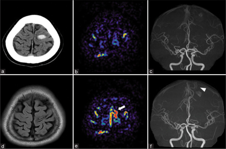
Figure 1:
(a) Computed tomography (CT) on admission. Magnetic resonance imaging (b and c) 3 days and (d-f) 93 days after onset (a) CT: Hematoma in the left primary motor cortex. (b) Arterial spin labeling (ASL): No obvious high signal around the hematoma. (c) Magnetic resonance angiography (MRA): No abnormal vessel around the hematoma (d) Flow-attenuated inverted recovery: Shrinkage of hematoma. (e) ASL: High focal signal on the convexity of the left frontal lobe and suprasagittal sinus (white arrow), which was not seen in previous ASL. (f) MRA showing a newly depicted vessel (white arrowhead) flowing in the direction of the hematoma.

