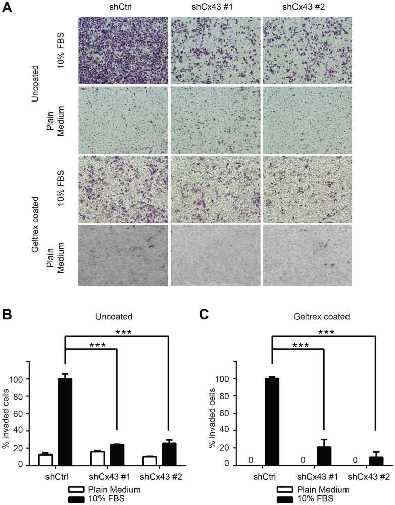
Figure 5. Cx43 is required for transwell invasion potential of PC-3 cells.
(A) Transwell invasion across uncoated (upper panel) and Geltrex coated (lower panel) inserts of control PC-3 cells and PC-3 cells with down-regulated Cx43 in the presence or absence of FBS, a chemoattractant. Cells invaded through the transwell membrane barrier were fixed and stained with crystal violet. Images were captured with a Leica DMI4000B microscope with a 10x objective lens. A representative microscopic field of each condition is shown. (B) Quantitation of the percentage of invaded cells across uncoated membrane. The invaded cells were quantified by determining the area of crystal violet staining using Image-J. The average area size from three independent microscopic fields was presented. Invasion of control shRNA transduced PC-3 cells in the presence of 10% FBS containing medium was used as reference. P values were calculated using one-way ANOVA and Dunnett’s post-test. *** P ≤ 0.001. (C) Quantification of the percentage of invaded cells across Geltrex coated membrane. Data were analyzed and plotted similarly as described in (B). *** P ≤ 0.001.

