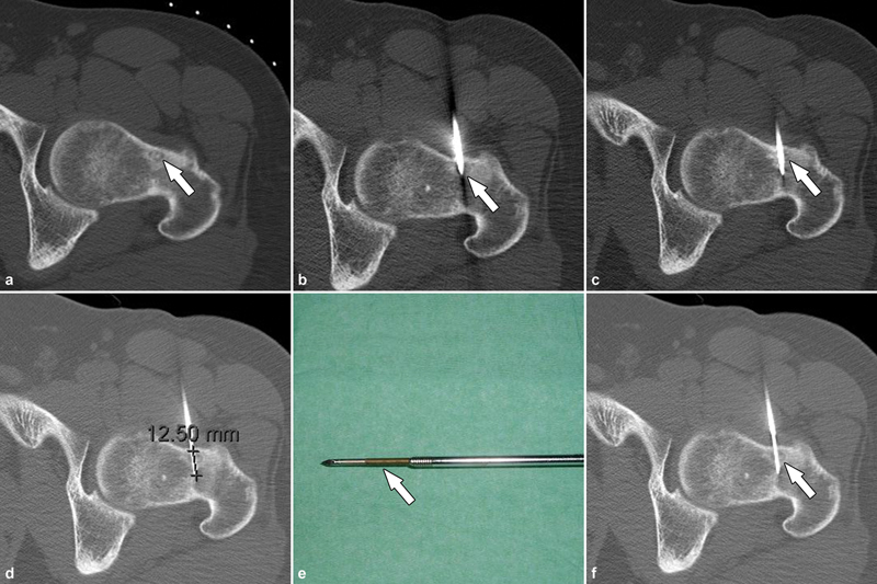
Fig. 4.

CT-guided percutaneous radiofrequency ablation under general anesthesia in supine position in a 25-year-old male presenting with left hip pain. ( a ) The sclerotic central nidus (arrow) is characteristically surrounded by a hypoattenuating halo and surrounding sclerotic reactive bone. ( b ) A 13-gauge coaxial bone drill system was utilized to penetrate the healthy cortical bone (arrow) to reach the central aspect of nidus ( c ). ( d ) To prevent unintentional supraphysiological heating of adjacent tissues along the tract of the trocar through heat conduction, the cannula with the trocar was partially withdrawn by ≥1 cm, while leaving the electrode tip ( e , arrow) in place before commencing ablation. ( f ) Once the electrode tip was located just beyond the central aspect of the nidus (arrow), ablation was initiated. As in this case, ablation often requires a single cycle lasting 4 minutes at 90 °C, but up to three treatment cycles may be performed depending on the size of the nidus to achieve a satisfactory ablation zone.
