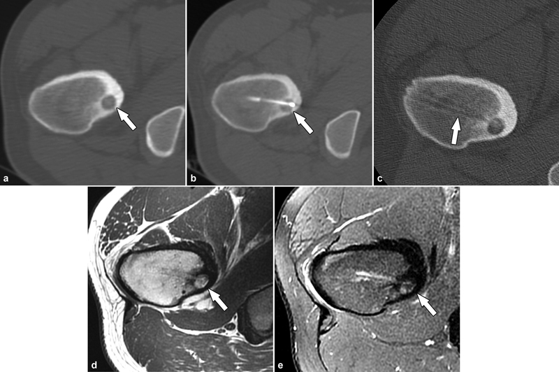
Fig. 5.

Percutaneous radiofrequency ablation of an osteoid osteoma in an 18-year-old patient. ( a ) Axial CT demonstrated the lucent nidus of the osteoid osteoma in the proximal femur (arrow) with surrounding reactive sclerosis. ( b ) Successful radiofrequency ablation of the nidus (arrow). ( c ) Final CT images demonstrated the needle track and the bony tunnel (arrow). Follow-up MRI at 12 months utilizing axial T1-weighted ( d ) and STIR ( e ) MR images demonstrated healing of the osteoid osteoma (arrows) and resolution of the bone marrow edema. The patient was pain-free postprocedure.
