
Fig. 26.
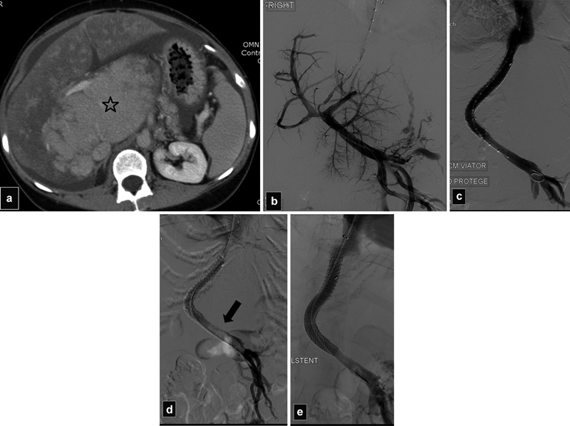
( a ) CT scan shows severe hypertrophy of the caudate lobe (star) and mottled enhancement of the liver in a patient with Budd–Chiari secondary to a hypercoagulable syndrome. ( b ) Initial portogram shows patent portal vein. ( c ) After TIPS with overlapping stents, the portal vein is patent. ( d ) Portogram the day after TIPS shows acute thrombosis of the main portal vein (arrow). ( e ) Portogram after declotting and additional stent placement into the main portal vein shows patent shunt.