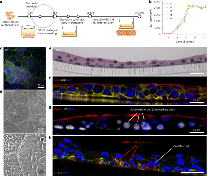
Fig. 1. The culture of USCs of juvenile C3H/HeN mice regenerates differentiated urothelium in vitro.
a, USCs isolated from 8-week-old C3H/HeN mice were expanded by spheroidal culture in matrigel with 50% L-WRN conditioned media (CM) including Y-27632, a ROCK inhibitor, and SB431542, a TGF β type 1 inhibitor. After 3 d of spheroid culture, cells were dissociated into a single-cell suspension and 3–4 × 10⁵ cells were seeded onto transwell membranes. The cells were cultured in 50% CM for 3–5 d, then cultured in 5% CM for 2–3 weeks until full differentiation. b, Cell cultures with a TER value >4,000 ohm × cm2 were then analysed (5 transwells cultured from one juvenile C3H/HeN cell line). For consistency, TER was measured 1 d after media change. c,d, Differentiated urothelia on the transwells were fixed and imaged via (c) confocal microscopy and (d) SEM to show a top-down view of the urothelium at magnification 500× (top panel) and 10,000× (bottom panel). In c, samples were stained for F-actin, the terminal differentiation marker K20 and nuclei (DAPI). e–h, The urothelia were also paraffin-embedded, sectioned and stained with hematoxylin and eosin (H&E) (e) and immunostained for K20, Ecad and DAPI (f), Upk3a, p63 and DAPI (g) or K5, K14 and DAPI (h). Representative images are shown. Data are from 2–3 independent experiments using USCs from 5 different juvenile C3H/HeN mice.

