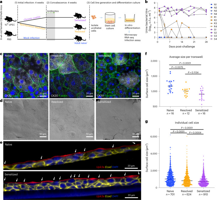
Fig. 2. Differentiated urothelia originating from previously infected mice maintain bladder remodelling phenotypes.
a, Time course of initial infection with 108 c.f.u. UTI89 KanR and convalescent period in C3H/HeN mice. b, Representative urine bacterial titre time course over 4 wpi. Horizontal line represents the cut-off for notable bacteriuria: 104 c.f.u. ml−1. Naïve, resolved and sensitized mice were named as N1-4, R1-4 and S1-4. c,d, USCs isolated from these mice were cultured into differentiated urothelia on transwells, fixed and imaged via confocal microscopy (c) and SEM (d). In c, the urothelia were stained for K20, F-actin (Phalloidin) and nuclei (DAPI). e, Transwells were paraffin-embedded, sectioned and immunostained for Upk3a, E-cadherin and nuclei. White arrows show cell junctions indicating size of surface cells. f,g, Fixed slides processed from 44 transwells of naïve, resolved and sensitized mice (n = 16, 12 and 16 transwells from n = 4, 3, 4 mice, respectively) were stained for K20, E-cadherin and nuclei, labelled and imaged in a double-blind manner. Then the superficial cell sizes were automatically measured using the Fiji ImageJ macro programme and plotted for average cell size per transwell (f) and individual cell size (g), represented as median with 95% CI. Two-tailed Student’s t-test was used to determine significance and P values are indicated when significant.

