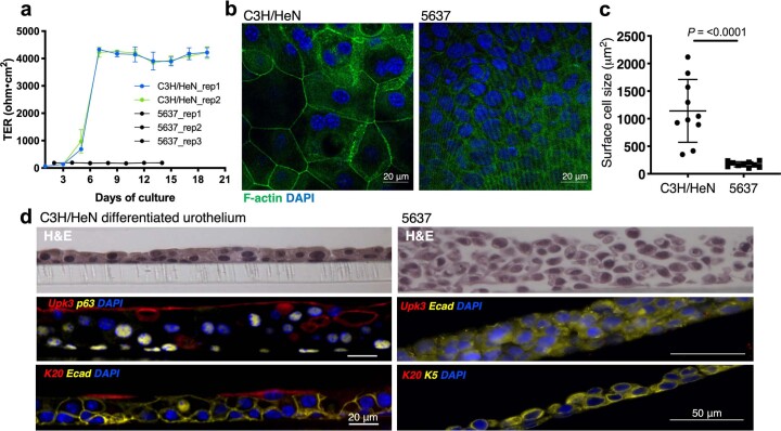
Extended Data Fig. 2. The differentiated urothelium better recapitulates bladder tissue phenotypes than the human bladder carcinoma cell line 5637 does, related to Fig. 1.
(A) Primary C3H/HeN urothelial cells and 5637 cells were cultured in Transwells for 2–3 weeks and transepithelial electrical resistance (TER) of Transwells were measured every 2 days before media change. Data collected from each cell lines (n = 3 for each) represented as mean ± SD. (B) Whole mount urothelium of both cell types were fixed and stained for confocal microscopy analysis; F-actin (green) and DAPI (blue). (C) Surface cell size of primary C3H/HeN urothelial cells and 5637 cells was measured using confocal images (n = 10 each). Data are represented as mean ± SD and significance was determined by an unpaired (two-tailed) t test (p-value <0.001). (D) The Transwell cultures of both C3H/HeN and 5637 cells were fixed, cut into slices, and then processed for paraffin embedding. Histologic sections were cut and stained with H&E or immunostained for Upk3a, K20, Ecad, K5, p63, and DAPI.

