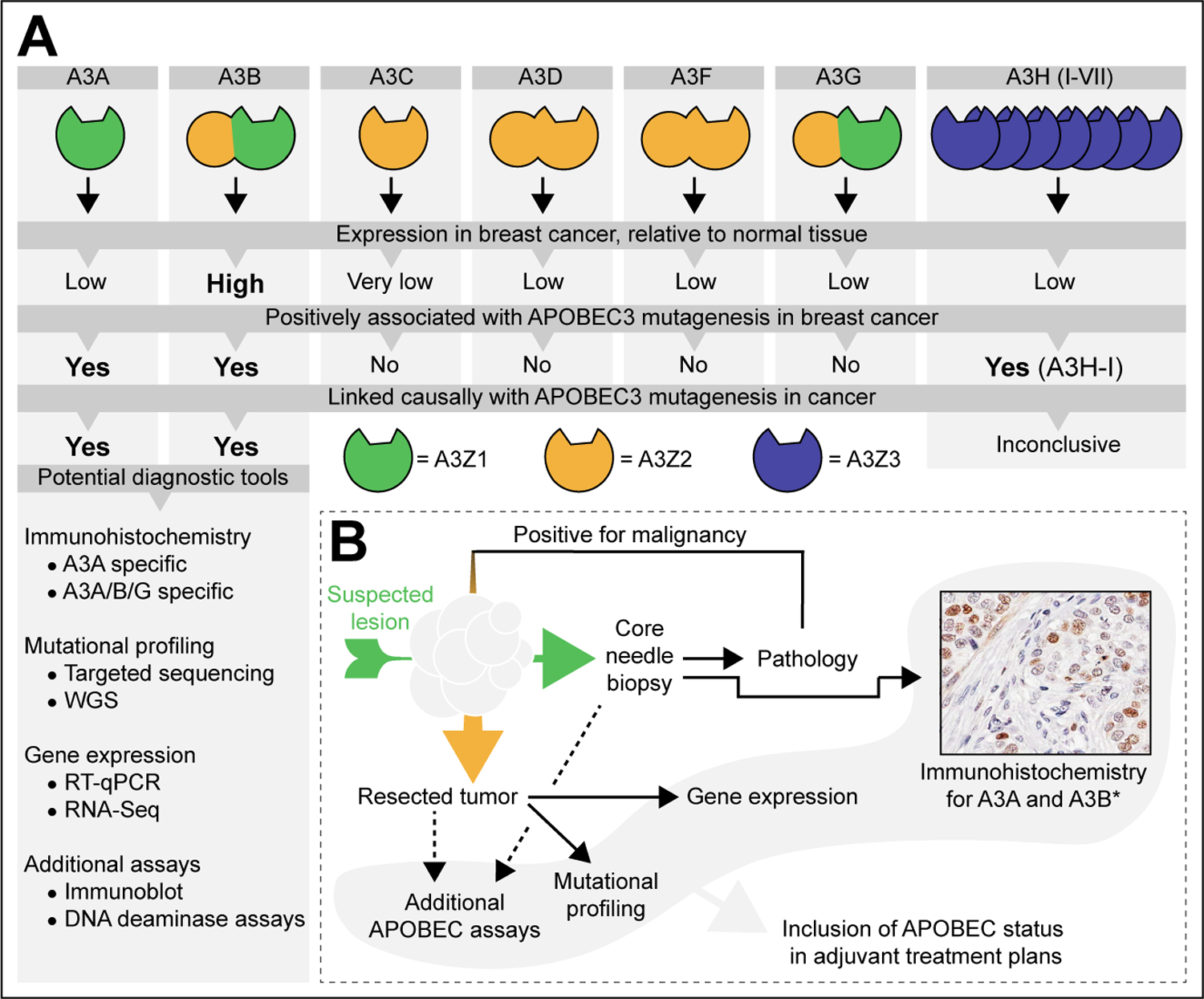
Figure 1. The APOBEC3 enzymes, their association with breast cancer, and the diagnostic methods available.

A Break-down of individual APOBEC3 family members, their conserved domain composition (5, 6), expression levels in cancer (17, 22, 30), and their causal involvement in the observed APOBEC mutagenesis pattern observed in cancer (13, 17, 22, 33). The list of potential diagnostic tools denotes published methods suitable for the detection of APOBEC3 enzymes, their deaminase activity, or the APOBEC single base substitutions [SBS] signatures (bioRxiv 2022.04.26.489523v2, (12, 17, 35, 39, 45–50).
B Proposed flow chart for the inclusion of the APOBEC status in the consideration of suitable adjuvant treatment plans. An initial core needle biopsy is taken from the suspected lesion [green arrow] and immunohistochemistry for A3A and/or A3B is performed in parallel to conventional clinical pathology. *The example shown here is considered A3B specific because of its nuclear localization. If malignant and operable, the freshly resected tumor [orange arrow] is subjected to additional assays, including mutational profiling and gene expression analyses. The resultant APOBEC status may then be included in the adjuvant treatment plans.
