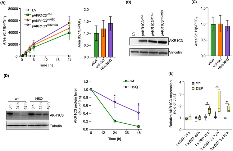
FIGURE 2.
The SNP rs12529 increases AKR1C3 protein stability and airborne PM upregulates AKR1C3 in human skin explants. A) LC–MS analyses of supernatants from HaCaT-AKR1C3-KO keratinocytes transfected with pAKR1C3wt, pAKR1C3H5Q, pAKR1C3wt/pAKR1C3H5Q (50:50), or empty vector. After 24 h, cells were treated with PGD2 (1 μM) for up to 24 h. Shown as mean ± SEM of n = 3 (left) and normalized to pAKR1C3wt/wt at 24 h (right). (B) AKR1C3 protein level of 24 h samples shown in (A). Representative blot of n = 3. (C) LC–MS quantification of 9α,11β-PGF2 after 24 h depicted in (A) normalized to AKR1C3 protein level shown in B). Shown as mean ± SEM of n = 3. (D) Keratinocytes were either transfected with pAKR1C3wt or pAKR1C3H5Q. After 24 h, cells were treated with 10 μM cycloheximide for up to 48 h. Representative blot (left) and mean quantification ± SEM of n = 4 (right). (E) Six μg/cm2 diesel exhaust particles (DEP) were topically applied to cultured human skin explants at days 1, 4, and 7. Skin samples were harvested after single, double, or triple DEP application for 3 days. Gene expression of AKR1C3 was analyzed by qPCR and normalized to 18 S rRNA level. *p ≤ 0.05.

