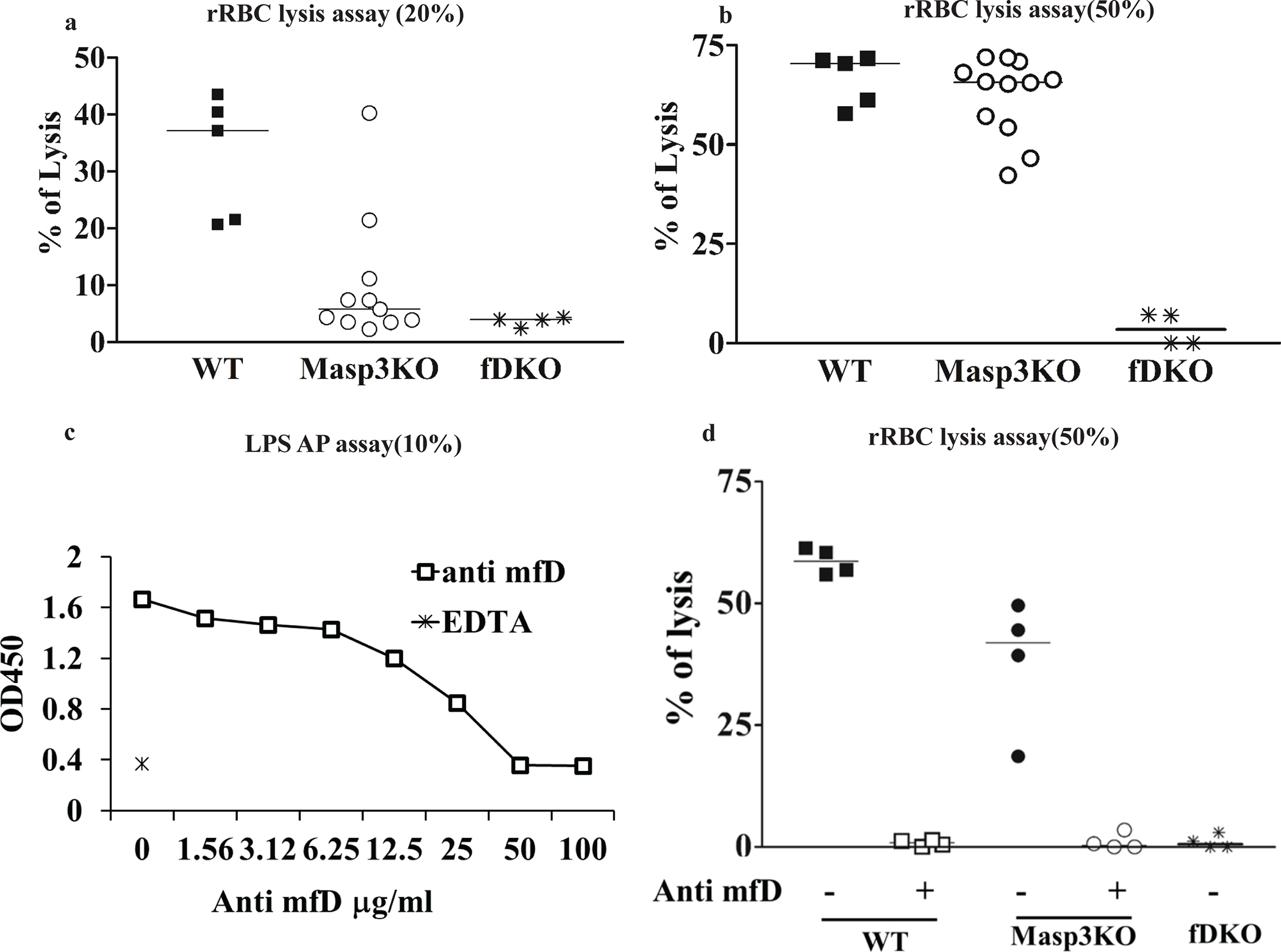
Fig 5: Assessment of AP complement activity in Masp3−/− mouse plasma using a rabbit red blood cell (RBC) hemolytic assay.

(a, b) Rabbit RBC lysis assays using 20% or 50% wildtype (WT), Masp3−/− (Masp3KO) or FD knockout (fDKO) mouse plasma. Rabbit RBC were easily lysed by wildtype mouse plasma but as expected, not by FD knockout mouse plasma. In contrast, significant lytic activity was detected in 20% plasma of some Masp3−/− mice, and close to wildtype mouse lytic activity was detected in 50% plasma of Masp3−/− mice. Each symbol represents data of plasma from an individual mouse. WT (n=5), Masp3KO (n=11) and fDKO (n=4). (c) Demonstration of dose-dependent inhibition of LPS-induced AP complement activity in 10% wildtype mouse plasma by a function-blocking mouse anti-mouse FD mAb (clone 14F11-3). At 50 or 100 μg/ml, mAb 14F11-3 completely inhibited mouse AP activity. EDTA serum was used as a positive control for complement inhibition. Values are average of duplicate assays using pooled plasma from 3–4 mice. (d) Effect of anti-FD mAb 14F11-3 (500 μg/ml) on rabbit RBC lysis by 50% wildtype (WT, n=4) or Masp3−/− (Masp3KO, n=4) mouse plasma. The data shows that AP complement activity in wildtype and Masp3−/− mouse plasma was completely blocked by mAb 14F11-3, suggesting that the AP activity detected in Masp3−/− mice was dependent on pro-FD and not on other nonspecific protease(s). FD knockout (fDKO) mouse plasma was used as a negative control which produced no hemolysis.
