
Figure 3: Potentiation of PVHTRH input to AgRP neurons is driven by PVHTRH neuron activity.
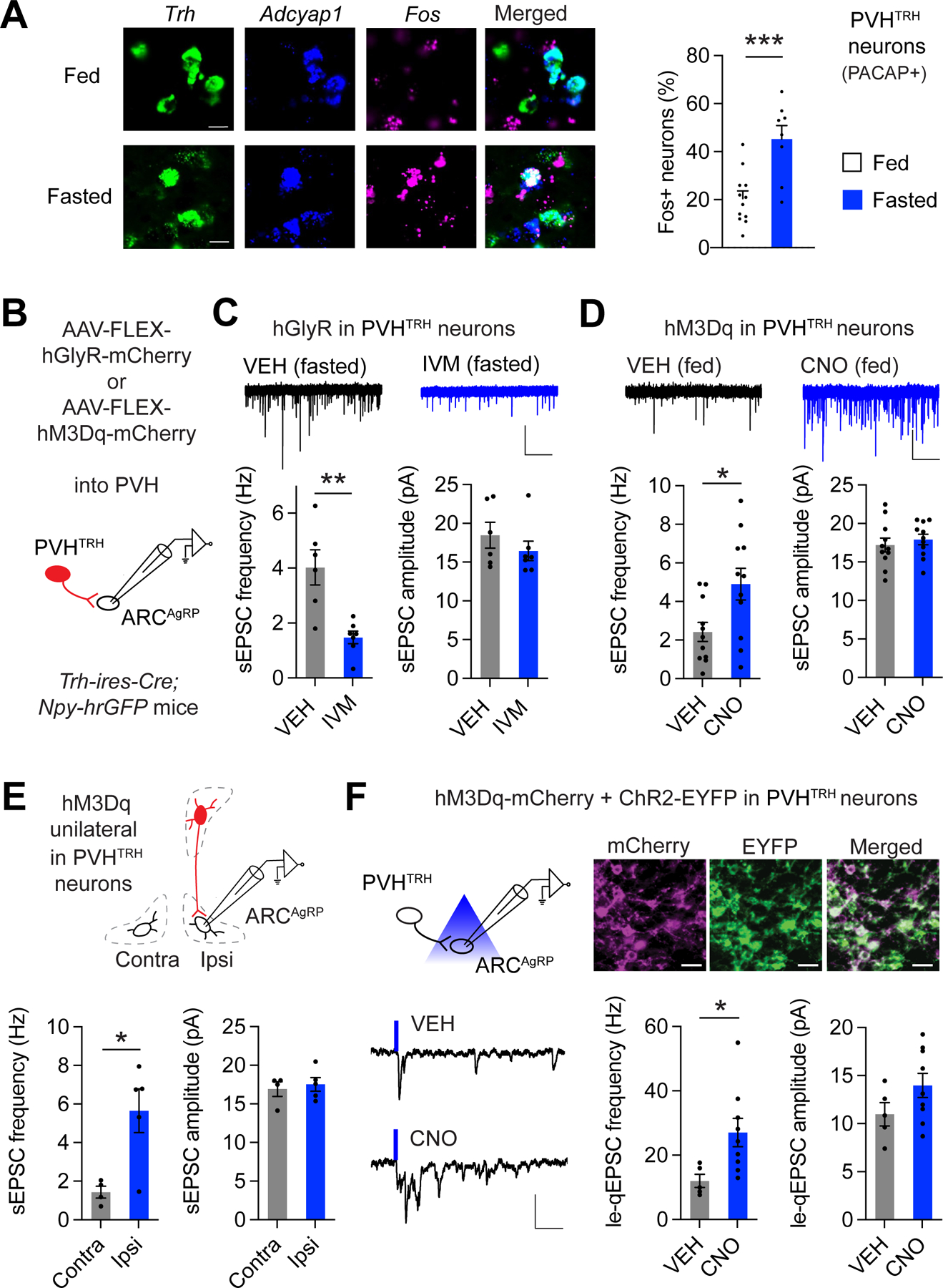
A) Representative histological images and analysis of Fos expression in PVHTRH neurons that express Adcyap1 (PACAP) from fed or fasted mice assessed by FISH. Scale bars represent 10 μm. (N = 4/4 mice).
B) Schematic of recordings from AgRP neurons following in vivo chemogenetic manipulations of PVHTRH neurons. Mice were injected with AAVs to express hGlyR or hM3Dq selectively in PVHTRH neurons.
C) Representative traces (top) of spontaneous excitatory postsynaptic currents (sEPSCs) in mice expressing hGlyR in PVHTRH neurons injected with VEH or IVM before fasting (scale bars: 25 pA, 2 s). Inhibition of PVHTRH neuron activity reduces the frequency, but not the amplitude, of sEPSCs in AgRP neurons in fasted mice (N = 2/2 mice).
D) Fed mice expressing hM3Dq in PVHTRH neurons were administered with vehicle (VEH) or CNO at the onset of the light cycle. Frequency, but not amplitude, of sEPSCs in AgRP neurons is significantly increased in mice treated with CNO 4 hours before brain slices are prepared (N = 2/2 mice; scale bar: 25 pA, 2 s).
E) Because chemogenetic stimulation of PVHTRH neurons acutely increases food intake (Figure S2B and 29), we tested the influence of circuit-specific activation on upregulation of sEPSC frequency. For this, hM3Dq was unilaterally expressed in PVHTRH neurons. In AgRP neurons recorded from the ipsilateral (Ipsi), but not from the contralateral (Contra), site of the stimulated PVHTRH neurons, sEPSC frequency is significantly upregulated, consistent with increased activity of the PVHTRH➔AgRP circuit as the functionally relevant mechanism for the amplification of excitatory drive onto AgRP neurons (N = 2/2 mice).
F) Schematic of the optogenetic approach to assess circuit-specific plasticity upon PVHTRH neuron activation (top left). Representative fluorescence images showing expression of ChR2-EYFP (green) and hM3Dq-mCherry (magenta) in the PVH (top right; scale bars, 20 µm). Note the co-expression of both viruses in PVHTRH neurons.
Representative traces (bottom left) of currents obtained from optical stimulation of PVHTRH afferents in presence of strontium from mice treated with VEH or CNO 4 hours before slice preparation (scale bars, 30 pA, 50 ms).
Summary of le-qEPSC recorded from AgRP neurons show that PVHTRH neuron activation significantly increases quantal frequency, but not amplitude, demonstrating that circuit activation amplifies activity of PVHTRH➔AgRP synapses (N = 5/4 mice).
All data are presented as mean ± s.e.m.; * p < 0.05, ** p < 0.01, ***p < 0.001; two-tailed unpaired Student’s t-test.